Delayed Onset Transient Diaphragmatic Paralysis after Pacemaker Implantation
Article information
Abstract
A 77-year-old woman presented with exertional dyspnea six days after left pectoral pacemaker implantation. Chest radiography at presentation showed that her left diaphragm was elevated when compared to earlier films. A fluoroscopic sniff test confirmed left diaphragmatic paralysis. Thoracic computed tomography did not reveal any major vascular or lung parenchymal injury; however, phrenic nerve injury on direct needle puncture during the original surgery was suspected. The patient’s small body size may have increased the risk of this injury. Delayed-onset unilateral diaphragmatic paralysis appears to be a rare complication of cardiac device implantation.
Introduction
Diaphragmatic paralysis can occur after cardiac surgery [1] or tumor growth [2]. It is also a rarely reported complication of cardiac device implantation [3,4]. We report a case of unilateral diaphragmatic paralysis due to complications during pacemaker implantation.
Case
A 77-year-old woman was admitted for exertional dyspnea. She was 148-cm tall and weighed 54 kg. The patient had undergone percutaneous coronary artery intervention 11 months ago for acute myocardial infarction with non–ST-segment elevation. Twenty-four hour Holter monitoring showed multiple episodes of paroxysmal atrial fibrillation followed by a prolonged sinus arrest over 10 seconds. It also detected persistent slow junctional escape rhythm lasting 2-3 hours. Paroxysmal atrial fibrillation with tachycardia-bradycardia syndrome was suspected. A dual-chamber pacemaker of the DDDR type was implanted for treatment. Venous angiography for the upper extremity showed normal axillary and subclavian vein anatomy. After local anesthesia with 1:1000 lidocaine, a subcutaneous pocket was created in the left pectoral area. The left subclavian vein was punctured and screw pacemaker leads (CapSureFix MRITM, Medtronic, Minneapolis, MN, USA) were inserted using the Seldinger method with guide wires and 8Fr peel-away introducers (Peel-Away IntroducerTM, St. Jude Medical, Minnetonka, MN, USA). Pacemaker leads were stably fixed to the right atrial appendage and apex within acceptable value of sensing and capture parameters. Diaphragm and pectoral muscle twitching were not induced by atrial and ventricular pacing at a high energy output (10 V×1.0 msec). Fluoroscopic examination performed immediately after pacemaker lead fixation did not show any evidence of diaphragmatic paralysis. The pacemaker leads and the generator were fixed to the left pectoral muscle. The procedure was finished in 90 minutes without complications. A postprocedural chest radiogram showed no abnormal findings such as pleural effusion or pneumothorax (Figure 1A). The patient was discharged 4 days later without exertional dyspnea.
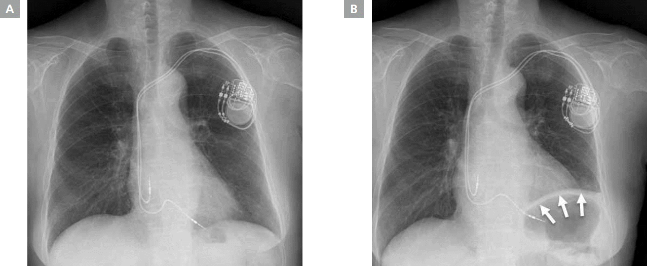
Chest radiography findings. (A) Chest radiogram obtained immediately after pacemaker implantation showed no abnormal findings such as pleural effusion or pneumothorax. (B) Chest radiogram obtained 10 days later showed elevation of the left diaphragm (white arrows).
Six days later, the patient presented with exertional dyspnea (New York Heart Association functional class II to III) and was readmitted. Her vital signs were stable and oxygen saturation level measured by arterial blood gas analysis was 97%. Twenty-four hour Holter monitoring showed an intermittent atrial back-up pacing rhythm without a significant sinus pause or junctional escape rhythm. The ejection fraction was 67% and there were no significant structural abnormalities found in echocardiographic examinations. Her chest radiogram showed elevation of the left diaphragm (Figure 1B), indicating left diaphragm paralysis. Fluoroscopic examination showed paradoxical movement of the left diaphragm during sniffing and deep inspiration. This indicated that the patient had paralysis on the left side of her diaphragm. Chest computed tomography was performed to determine the cause of diaphragmatic paralysis. There was no hematoma detected at the left subclavian vein puncture site or postulated course of the left phrenic nerve. There was no evidence of cardiac perforation, pericardial effusion, or pneumothorax (Figure 2). The pacemaker was operating under normal parameters. Prednisolone (0.5 mg/kg/day) was administered for 5 days but dyspnea did not resolve. Pulmonary function testing showed decreased lung function of a restrictive pattern. The patient had a 53% forced expiratory volume in one second and a 52% forced vital capacity. The respiratory distress was not severe, so the expectation was that the diaphragmatic paralysis would spontaneously recover. The patient was discharged with a furosemide prescription and the establishment of a physical exercise program. After six months, the patient’s symptoms resolved and diaphragmatic paralysis recovered spontaneously without surgical intervention.

Chest computed tomography findings. Black arrows show the course of pacemaker leads from the subclavian vein to the cardiac chambers. (A, B) No hematoma was detected at the subclavian vein puncture site or the postulated course of the left phrenic nerve. (C, D) There was no evidence of cardiac perforation by the pacemaker leads, pericardial effusion, or pneumothorax. (D) Chest computed tomography showed elevation of the left diaphragm (white arrows) with subsegmental atelectasis.
Discussion
The diaphragm is a major respiratory muscle innervated by phrenic nerves. The phrenic nerves originate from the cervical spinal root (C3 to C5). These nerves ordinarily enter into the thoracic cavity between the subclavian artery and vein. They then travel to the diaphragm region along the pericardiophrenic vessels and lie between the pericardium and parietal pleura in the thoracic cavity [5,6].
A number of diseases such as inflammatory diseases and surgeries such as cardiac surgery, thoracic tumor, subclavian or internal jugular vein cannulation, and radiofrequency catheter ablation cause phrenic nerve injury, which may lead to early- or delayed-onset diaphragmatic paralysis [7–12]. Paralysis of the diaphragm can be easily diagnosed by a fluoroscopic sniff test. However, it is not always possible to determine the precise cause of paralysis. Phrenic nerve paralysis from cardiac device implantation has rarely been reported in the literature [3,4]. Phrenic nerve compression has been caused by traumatic internal mammary artery pseudoaneurysm. Indirect phrenic nerve injury via electrocautery or local anesthesia has also been reported.
In the present case, phrenic nerve injury by subclavian vein puncture needling appears highly probable, given the patient’s small body size and a lack of evidence for major vascular or lung parenchymal complications on chest computed tomography. Phrenic nerves enter the thoracic cavity near the internal jugular and subclavian vein junction. This region is smaller in patients with smaller bodies, which increases the risk of direct phrenic nerve injury on subclavian vein puncture needling. Several phrenic nerve anatomic variations have been reported to be susceptible to direct needle puncture injury. For example, the location where the ipsilateral phrenic nerve penetrates the subclavian vein is susceptible to direct needle puncture injury [13].
Previous case studies have reported immediate-onset dyspnea after device implantation. However, dyspnea began 6 days after pacemaker implantation in this case. There were no symptoms or signs indicating diaphragmatic paralysis on chest radiography or fluoroscopic examination performed immediately after pacemaker implantation. The cause of delayed onset diaphragmatic paralysis may be related to Wallerian degeneration of the damaged nerve, which reportedly takes one week to three months.
Microhematoma (undetected by chest CT) may also have caused phrenic nerve compression as the patient was administered dual antiplatelet agents at the time of the procedure. In addition, direct or indirect phrenic nerve injuries by relatively stiff pacemaker leads or peel-away introducers may be other causes of diaphragmatic paralysis. Inflammatory or thrombotic changes of the punctured left subclavian vein may have influenced the adjacent left phrenic nerve. There is currently no consensus on the treatment of diaphragmatic paralysis. Fortunately, respiratory distress was not severe in this patient as there was no underlying lung parenchymal disease. Asymptomatic unilateral diaphragmatic paralysis generally spontaneously resolves in one to two years. However, surgical diaphragmatic plication may be necessary for severely symptomatic cases.
Diaphragmatic paralysis is a rare complication of cardiac device implantation. Phrenic nerve injury may be caused by subclavian puncture needling during the device implantation procedure. Surgeons should consider the complex anatomy of the phrenic nerve when performing cardiac device implantation. This is particularly important for patients with a relatively small body size. The surgeon needs to minimize the risk of phrenic nerve injury by using fluoroscopic guidance during axillary vein puncture.
Acknowledgements
The authors declare no financial conflicts of interest.