Introduction
Outflow tracts are common sites of origin for ventricular tachycardia (VT) or a ventricular premature complex (VPC), especially in young patients without structural heart disease. Up to 90% of outflow tract VT is considered to originate from the right side, mainly from the right ventricular outflow tract (RVOT) [1]. Left ventricular outflow tract (LVOT) tachycardia can arise from any aspect of the heart, including endocardial or epicardial sites [2]. The origin of idiopathic outflow tract VT can be closely related to important adjacent structures like the aortic valve leaflet and coronary artery; therefore, radiofrequency (RF) ablation can damage these structures without an understanding of the anatomical relationships [3,4]. In this respect, intracardiac echocardiography (ICE) can improve the safety and efficacy of RF ablation by identifying anatomical details not visible by fluoroscopy and by confirming stable catheter placement and lesion formation at the target site [5,6].
Case
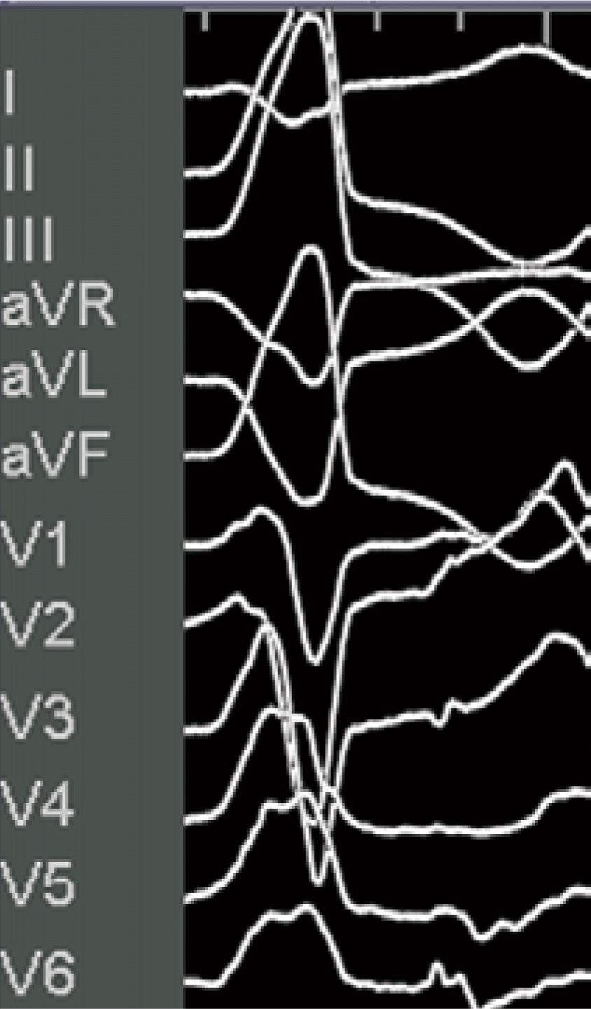
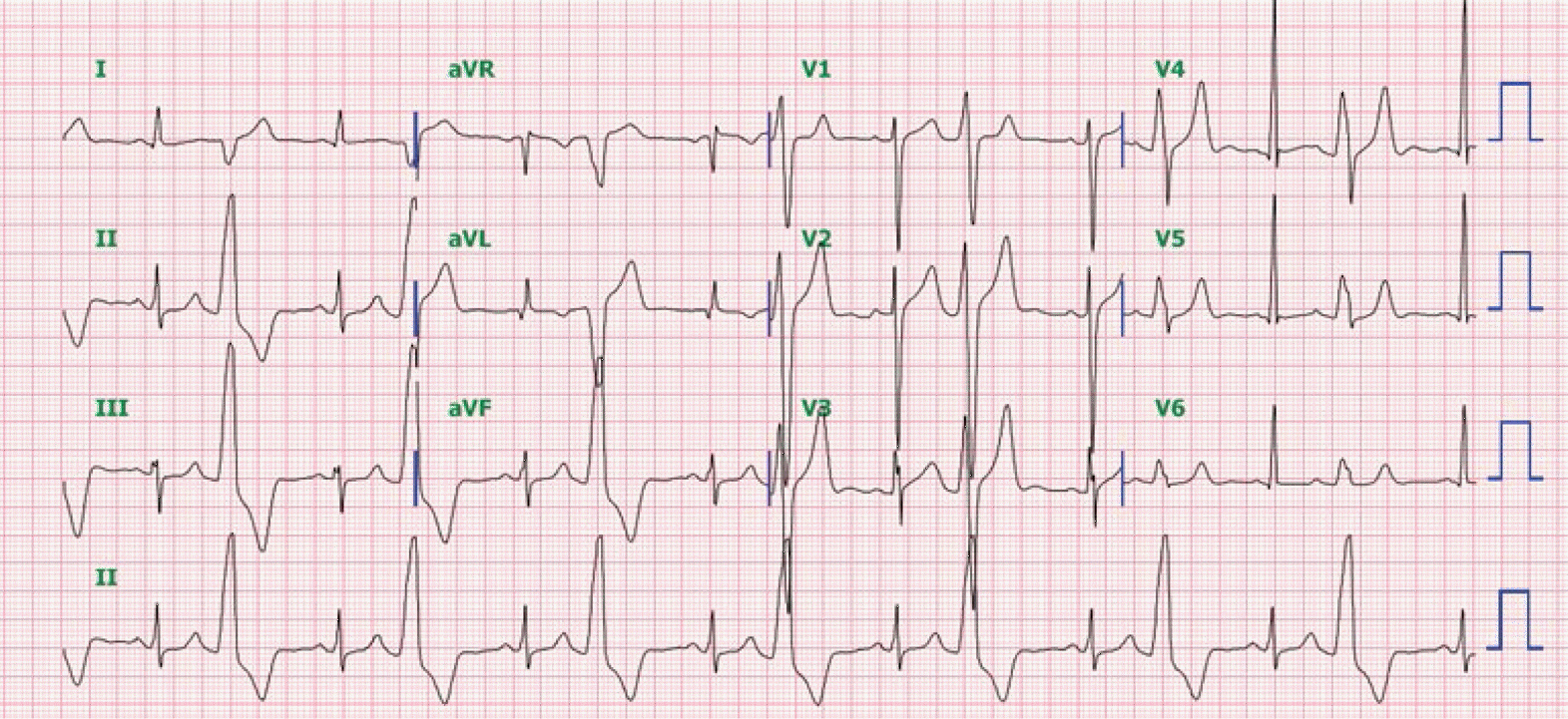
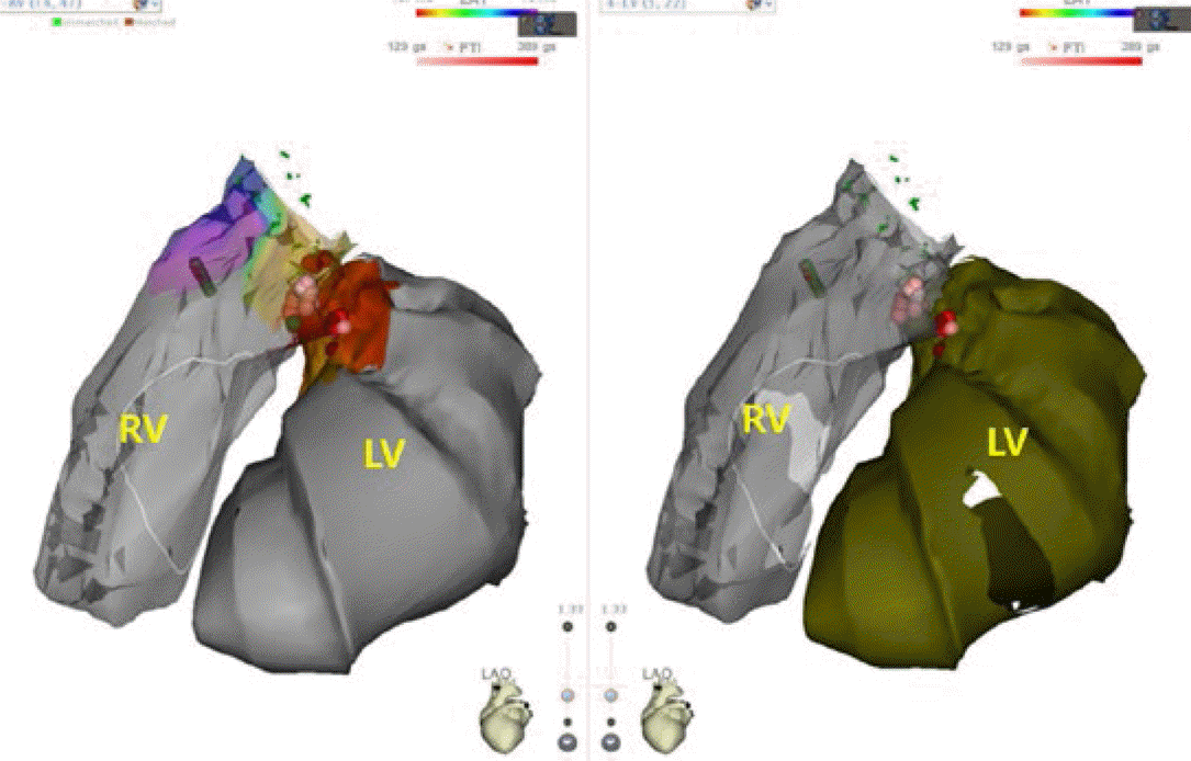
A 40-year-old man referred to our hospital presented with palpitations and dyspnea. The patient had been followed-up for a year, with frequent monomorphic VPCs at a local clinic. He had no underlying medical conditions or structural heart disease at the time of diagnosis. Trans-thoracic echocardiography was performed at our hospital one year after diagnosis, and it showed moderate left ventricular (LV) systolic dysfunction with an LV ejection fraction less than 35%. Twenty four-hour Holter monitoring revealed a VPC burden of 34% and episodes of non-sustained ventricular tachycardia. RF ablation was indicated for the patient because of symptomatic VPC with LV dysfunction. Surface electrocardiogram (ECG) showed VPC bigeminy with a left bundle branch block pattern, inferior axis, precordial transition of V3, and Q wave in lead I (Figure 1). RF energy (maximum 30W, 60 seconds per each ablation) was delivered at the RVOT anterior septum. We also mapped the LVOT septal area both above and below the right and left coronary cups, because there was no response after two applications of RF energy in the RVOT area. Discrete potentials preceding 81 milliseconds before the local ventricular electrogram were noted in the LVOT septum below the left and right coronary cusp junction opposing the RVOT ablation site (Figure 2). A total of 27 RF energy applications were made at this site and VPC were observed very seldom after the procedure. One month after the procedure, improvement in symptoms and LV systolic function (ejection fraction, 47.2%) was noted. However, follow-up ECG showed recurrent VPC bigeminy with a large VPC burden (26%) on follow-up 24-hour Holter monitoring. We considered the probability of VPCs originating from the LV summit, because a precordial pattern break was present, which showed a smaller R wave in lead V2 than in V1 and V3, and maximum deflection index >55% in 12-lead ECG before the second procedure (Figure 3).
Figure 2.
Three-dimensional mapping images by CartoUnivu system showing ablation lesions in the right and left outflow tract septa during the first radiofrequency ablation procedure
LVOT, left ventricular outflow tract; RVOT, right ventricular outflow tract

Figure 3.
Recurrent ventricular premature complex on 12-lead electrocardiogram after the first procedure

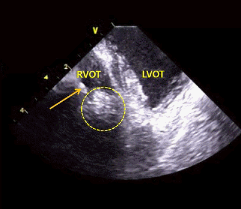
The redo procedure was planned under the guidance of ICE. Although the possibility of VPCs originating from the LV summit was apparent, we began mapping the outflow tract endocardium first based on the previous report showing successful ablation of LV summit VT from the opposite endocardial side in more than half of the cases [7]. We ablated the earliest activation site with a good pacemap in the RVOT anterior septum and confirmed stable contact of the catheter to the ablation site by ICE. Due to the occasional appearance of VPCs, we mapped the LVOT area additionally by trans-aortic approach. The earliest activation site was directly opposite to the RVOT ablation lesions. During the application of RF energy (maximum 40W, 160 seconds), stable contact of the catheter was confirmed by ICE. Lesion formation was also clearly visualized by ICE and the transmural lesion was identified (Figure 4A and 4B). Unlike in the first ablation procedure, VPCs disappeared within a second of application of RF energy and no VPC appeared until discharge. At the 1-month follow-up, the patient no longer complained of palpitations and no single VPC was seen on 12-lead ECG.
Discussion
Structurally, the outflow tracts are common sites for the origin of VT or VPC in the normal heart [1,2]. Although outflow tract VT prognosis is favorable, there is potential for VPC-related cardiomyopathy or even sudden cardiac arrest [8,9]. Catheter ablation is an effective therapeutic option as well as medical therapy such as a calcium channel blocker [3,10]. However, it is very important to understand the anatomical relationships between the outflow tracts and adjacent cardiac structures to safely perform catheter ablation. The LV summit is defined as the LV epicardial surface bounded by the left anterior descending and left circumflex arteries, which is the most superior part of the LV [2]. The endocardial LV below the left coronary cusp represents the opposite aspect of the LV summit. LV summit VT typically has a right bundle branch block pattern, but a left bundle branch block pattern with inferior axis and early transition can also be present. An epicardial origin is suggested by ECG characteristics including a pseudo-delta wave ≥34 ms, pattern break in the precordial leads, and maximum deflection index ≥0.55 [11]. Because of its proximity to major coronary arteries and fat tissue, a direct epicardial approach is usually limited. Therefore, if ablation within the coronary venous system is not feasible or safe, an attempt can be performed from the LV endocardium. If this approach is insufficient regarding effectiveness, ablation from the RVOT septum can be attempted with a longer application of RF energy with higher power [12].
ICE is an excellent tool for visualizing the anatomical relationship and endocardial details that are not visible using fluoroscopy. It provides real-time imaging, which enables confirmation of catheter contact or lesion formation, and early detection of complications such as tamponade [4–6]. It can also help to reduce the risk during ablation of potentially dangerous sites such as near coronary vasculatures or valve leaflets. In addition, higher energy and RF for longer duration can be delivered during ablation, avoiding complications because of ablation catheter placement under direct visualization, and lesion formation can be directly visualized. In summary, ICE can improve the safety and efficacy of RF ablation, especially at sites with complex anatomical relationships or an intramural origin.


